

Contact us
E.mail : infos@eyecare.com
Address : 1 Dmshek,Maadi, Cairo
Eye Care : Sat. - Thu. 9:00am - 10:00pm

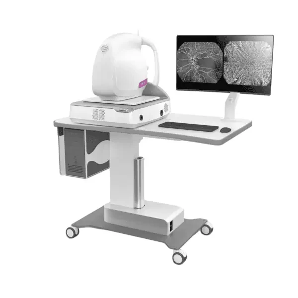
OCT – Optical Coherence Tomography
Retinal & Macular OCT and OCT Angiography (OCTA)
Optical Coherence Tomography (OCT) is an essential imaging technique used in the diagnosis and monitoring of many retinal and optic nerve conditions. It provides detailed cross-sectional images of the retinal layers, helping ophthalmologists identify structural changes that may not be visible during a routine fundus examination.
At the Eye Scan Unit of Eye Care Center, these examinations are performed using the TowardPi YAlkaid imaging system. The device combines advanced retinal and fundus imaging technologies to provide detailed information that supports diagnosis, disease monitoring, and treatment assessment.
Available Imaging Examinations
1. Retinal and Macular OCT
Provides detailed cross-sectional images of the retinal layers and macula. It helps diagnose and monitor conditions such as:
-
Macular edema and swelling.
-
Diabetic retinopathy.
-
Age-related macular degeneration (AMD).
-
Macular holes and epiretinal membranes.
-
Other conditions affecting retinal structure.
2. Optic Nerve OCT
Evaluates the optic nerve and the surrounding retinal nerve fiber layer. It is commonly used in the assessment and monitoring of glaucoma and other conditions that may affect the optic nerve.
3. OCT Angiography (OCTA)
An advanced imaging technique that visualizes the retinal and macular blood vessel networks by detecting the movement of blood cells within the vessels, without the need for dye injection. It helps assess vascular changes associated with selected retinal conditions.
4. Multicolor Fundus Imaging
Uses different wavelengths of light to produce detailed fundus images, highlighting different retinal structures and supporting the assessment and monitoring of certain retinal conditions.
5. Fundus Autofluorescence (FAF)
Helps evaluate changes associated with the retinal pigment epithelium and surrounding retinal tissues. It is particularly useful in the assessment and monitoring of selected retinal disorders affecting these structures.
Benefits of OCT and OCTA Imaging
These imaging techniques help ophthalmologists to:
-
Obtain detailed images of retinal layers and the optic nerve.
-
Detect and monitor structural changes associated with retinal diseases.
-
Assess macular edema before and after treatment.
-
Monitor selected glaucoma cases and changes affecting the optic nerve.
-
Visualize retinal microvasculature using OCTA without dye injection.
The ophthalmologist determines the appropriate examination based on the patient's symptoms, clinical findings, and medical condition. The required imaging tests may vary from one patient to another.



Connect Us