

Contact us
E.mail : infos@eyecare.com
Address : 1 Dmshek,Maadi, Cairo
Eye Care : Sat. - Thu. 9:00am - 10:00pm


Ultrasonography
Ocular and Orbital Ultrasound
Ocular and orbital ultrasound is an important diagnostic imaging examination in ophthalmology. It helps assess the internal structures of the eye and surrounding orbital tissues, particularly when direct examination of the retina and fundus is difficult or impossible.
The examination uses high-frequency sound waves to produce images and measurements of the internal structures of the eye, helping ophthalmologists diagnose and monitor various eye conditions.
Types of Ocular Ultrasound
Eye Care Center provides two types of ocular ultrasound examinations:
1. A-Scan (Amplitude Scan)
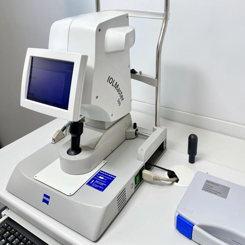
A-Scan is used to measure the dimensions of the eye, particularly its axial length. It is an important measurement for calculating the appropriate power of the artificial intraocular lens before cataract surgery.
2. B-Scan (Brightness Scan)
B-Scan produces a two-dimensional image of the internal structures of the eye. It helps assess the retina and vitreous and detect various conditions, particularly when ocular opacity prevents direct visualization of these structures.
When Is an Eye Ultrasound Needed?
An ophthalmologist may recommend this examination in several situations, including:
-
Dense Cataracts: When severe lens opacity prevents clear visualization of the retina and fundus, ultrasound helps assess the posterior segment of the eye.
-
Vitreous Hemorrhage: Helps evaluate the retina and detect possible retinal tears or detachment.
-
Retinal Detachment: Helps identify retinal detachment and assess its extent.
-
Intraocular Tumors: Helps evaluate certain tumors inside the eye, including their characteristics and extent.
-
Orbital Conditions: Helps assess certain abnormalities and masses affecting the tissues surrounding the eye.
Ocular and orbital ultrasound provides valuable diagnostic information that helps the ophthalmologist determine the appropriate management plan based on the patient's condition and other examination findings.


Connect Us